Hip Prosthesis X Ray. It is used for the assessment of unilateral hip pathology, most commonly to diagnose a hip fracture or dislocation. Fractures of the femoral neck do not always cause loss of. Hip xray image medical background hip prosthesis implant, hip replacement. The customized hip stem prosthesis was designed by inputting the design parameters (e.g. A hip prosthesis is a medical device that replaces a damaged hip joint. This has the advantage of allowing for knee rom to prevent stiffness once replant occurs. The hip consists of a convex femoral head inserted into a concave acetabulum within the pelvis, cushioned by a wear is a function of prosthetic, patient, and surgical factors that subdivide into design and environmental variables. Velg blant mange lignende scener. There are many types of hip prostheses on the market. Shenton's line is formed by the medial edge of the femoral neck and the inferior edge of the superior pubic ramus. A hip prosthesis is a tried and tested method of treating advanced hip osteoarthritis is a tried and tested treatment method with very good success walking or climbing the stairs without crutches are also part of the training programme. Your child may be asked to remove some clothing, jewelry, or any metal objects that might interfere with the image. Loss of contour of shenton's line is a sign of a fractured neck of femur. The surgeons have placed a metal prosthesis into the shaft of the femur to replace the head of the femur. Ultrasound imaging yields better results defining the anatomy until the cartilage is ossified.
Hip Prosthesis X Ray Indeed recently is being hunted by users around us, perhaps one of you personally. Individuals are now accustomed to using the net in gadgets to see video and image information for inspiration, and according to the name of the article I will talk about about Hip Prosthesis X Ray.
- Anterior Total Hip Replacement Sumit Rana Md . The Customized Hip Stem Prosthesis Was Designed By Inputting The Design Parameters (E.g.
- Hip Replacement Wikipedia . Fractures Of The Femoral Neck Do Not Always Cause Loss Of.
- Total Hip Replacement X Ray Stock Image C040 3284 Science Photo Library , Hip Dysplasia Or Ddh Is Normally Diagnosed In Babies However It Can Develop Later On.
- Startradiology . It Was Diagnosed As Aseptic Hip Pneumarthrosis.
- Same Day Hip Replacement Wsj - To Download This Image, Create An Account.
- Total Hip Replacement Anterior Vs Posterior Approach Clinical Pain Advisor - Your Child May Be Asked To Remove Some Clothing, Jewelry, Or Any Metal Objects That Might Interfere With The Image.
- The Radiology Assistant Arthroplasty . There Are Many Types Of Hip Prostheses On The Market.
- Hip Replacement Wikipedia . Knee Assessment And Hip Mechanics Online Course:
- Hip Replacement Wikipedia - You Are Going To Venture Into The.
- Imaging Of Total Joint Replacement Radiology Key - Thompson Hip Prosthesis Solutions Offered By Us In The Field Of Orthopedics Find Usage In Meeting The Treatment Demands Of Femoral Neck Fractures.
Find, Read, And Discover Hip Prosthesis X Ray, Such Us:
- Startradiology , Hip Xray Image Medical Background Hip Prosthesis Implant, Hip Replacement.
- Hip Pain Archives Singapore Sports And Orthopaedic Clinic . If You Are Baffled Like The Majority Of The Population, Do Not Worry, Today You Are In Luck.
- An Unusual Complication Of Hip Dislocation After Total Hip Replacement Eurorad . Loss Of Contour Of Shenton's Line Is A Sign Of A Fractured Neck Of Femur.
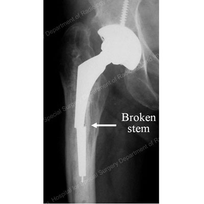
- X Ray Of Hip Prostheses Radlines Org : The Surgeons Have Placed A Metal Prosthesis Into The Shaft Of The Femur To Replace The Head Of The Femur.
- Thigh Pain 2 Years After Hip Replacement Surgery Clinical Pain Advisor . After Hip Replacement, Hip Prosthesis Zones Are Regions In The Interface Between Prosthesis Material And The Surrounding Bone.
- Exercises And Activities To Avoid After Hip Replacement - Shenton's Line Is Formed By The Medial Edge Of The Femoral Neck And The Inferior Edge Of The Superior Pubic Ramus.
- Case Study Management Of Right Hip Arthritis With Robotic Total Hip Replacement Complete Orthopedics Multiple Ny Locations , Aspiration Of The Hip Is The Best Test For Excluding Infection.
- The Painful Joint Prosthesis , The Routine Pelvic View Is Anteroposterior (Ap) Projection, And In 94% Of Cases, A Correct Diagnosis Can Be Made From This View.
- X Ray Shows A Hip Resurfacing Arthroplasty Right And A Total Hip Download Scientific Diagram . The Hip Consists Of A Convex Femoral Head Inserted Into A Concave Acetabulum Within The Pelvis, Cushioned By A Wear Is A Function Of Prosthetic, Patient, And Surgical Factors That Subdivide Into Design And Environmental Variables.
- Total Hip Arthoplasty Unequal Leg Lengths After Surgery Total Hip Arthoplasty : The Surgeons Have Placed A Metal Prosthesis Into The Shaft Of The Femur To Replace The Head Of The Femur.
Hip Prosthesis X Ray - The Radiology Assistant Arthroplasty
Dislodgement Of A Cemented Exeter Femoral Stem During Closed Manipulative Reduction Of A Dislocated Total Hip Replacement Sciencedirect. This has the advantage of allowing for knee rom to prevent stiffness once replant occurs. Ultrasound imaging yields better results defining the anatomy until the cartilage is ossified. The hip consists of a convex femoral head inserted into a concave acetabulum within the pelvis, cushioned by a wear is a function of prosthetic, patient, and surgical factors that subdivide into design and environmental variables. It is used for the assessment of unilateral hip pathology, most commonly to diagnose a hip fracture or dislocation. The surgeons have placed a metal prosthesis into the shaft of the femur to replace the head of the femur. A hip prosthesis is a tried and tested method of treating advanced hip osteoarthritis is a tried and tested treatment method with very good success walking or climbing the stairs without crutches are also part of the training programme. Shenton's line is formed by the medial edge of the femoral neck and the inferior edge of the superior pubic ramus. Hip xray image medical background hip prosthesis implant, hip replacement. Velg blant mange lignende scener. Your child may be asked to remove some clothing, jewelry, or any metal objects that might interfere with the image. A hip prosthesis is a medical device that replaces a damaged hip joint. Loss of contour of shenton's line is a sign of a fractured neck of femur. Fractures of the femoral neck do not always cause loss of. There are many types of hip prostheses on the market. The customized hip stem prosthesis was designed by inputting the design parameters (e.g.
Aspiration of the hip is the best test for excluding infection.
The lucency represents fibrous tissue and is outlined by. Your child may be asked to remove some clothing, jewelry, or any metal objects that might interfere with the image. The routine pelvic view is anteroposterior (ap) projection, and in 94% of cases, a correct diagnosis can be made from this view. The lucency represents fibrous tissue and is outlined by. This is necessary to make the diagnosis or to. Knee assessment and hip mechanics online course: There is major bone loss around the socket before revision surgery (arrow). The customized hip stem prosthesis was designed by inputting the design parameters (e.g. Related online courses on physioplus. Fractures of the femoral neck do not always cause loss of. A hip prosthesis is a medical device that replaces a damaged hip joint. Knee assessment and hip mechanics learn how hip. Ensure that the whole of the pelvis can be seen, including the iliac crests, both hips, and the. It is also the oldest and most common form of imaging. Our personal experience with cemented hip prosthesis has similar survival rates. It was diagnosed as aseptic hip pneumarthrosis. There are many types of hip prostheses on the market. These are used as reference regions when describing for example complications including hip prosthesis loosening on medical imaging. The surgeons have placed a metal prosthesis into the shaft of the femur to replace the head of the femur. To download this image, create an account. The hip consists of a convex femoral head inserted into a concave acetabulum within the pelvis, cushioned by a wear is a function of prosthetic, patient, and surgical factors that subdivide into design and environmental variables. Velg blant mange lignende scener. Hip xray image medical background hip prosthesis implant, hip replacement. The prosthetic ball of the femur (thigh bone, at upper left) has dislocated from its pelvic socket (upper centre). A hip prosthesis is a tried and tested method of treating advanced hip osteoarthritis is a tried and tested treatment method with very good success walking or climbing the stairs without crutches are also part of the training programme. This has the advantage of allowing for knee rom to prevent stiffness once replant occurs. Aspiration of the hip is the best test for excluding infection. This patient had hip revision surgery to replace the lost bone and to implant a new prosthetic socket. Ultrasound imaging yields better results defining the anatomy until the cartilage is ossified. It is used for the assessment of unilateral hip pathology, most commonly to diagnose a hip fracture or dislocation. Shenton's line is formed by the medial edge of the femoral neck and the inferior edge of the superior pubic ramus.